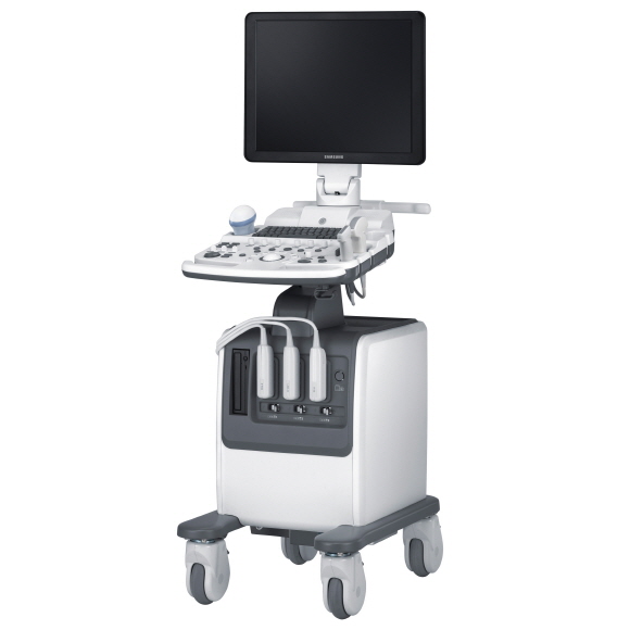
SonoAce R7
Ultra-Compact Unit with Advanced Performance
TGet outstanding 2D color performance and a simple user interface with this system,
ensuring an increase in patient output. It is ergonomic and offers sensitive waves and Color Doppler.
Features
The ergonomic and ultra-compact SonoAce R7 increases clinical efficiency through its combination of a simple user interface. It includes features such as second-stage filtering and spatial compounding to reduce extraneous noise and speckle while producing crystal-clear, high resolution results. By utilising outstanding Samsung 3D/4D color technology, images are easily viewed on a 19-inch LCD monitor. Clinical productivity is enhanced by customisable menus and a user-friendly design. A suite of extended tools ensures practitioners to have a wide range of resources to deliver timely, comprehensive and reliable diagnoses across multiple applications.
Slim and Compact Design
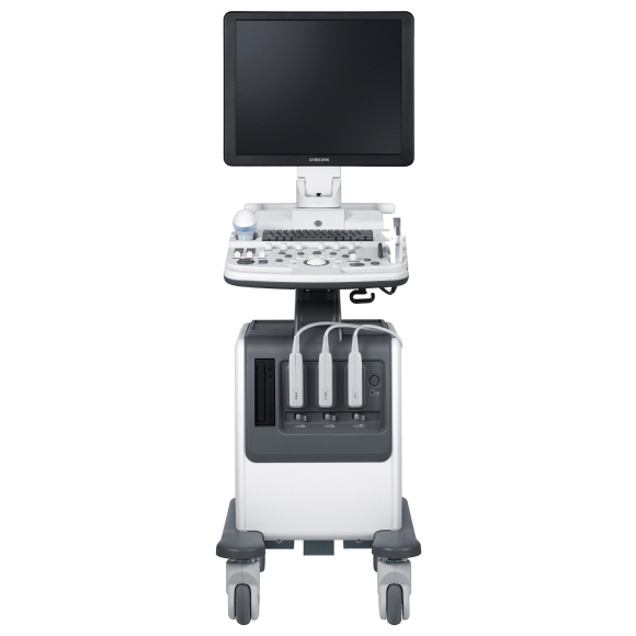
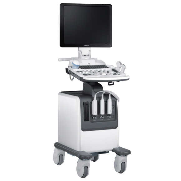
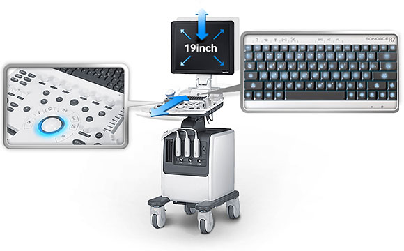
The user-friendly SonoAce R7 incorporates a host of ergonomic features into an ultra-compact package. The slim design is also practical with its four swivel wheels and front and back handles enabling easy mobility. The simple user interface, height-adjustable control panel and backlit keyboard combine to create a tool which enables practitioners to increase patient throughput without sacrificing comfort or performance. By utilising outstanding Samsung 3D/4D color technology, its 19-inch LCD monitor displays clear, easily-viewable images to bring about increased diagnostic accuracy.
Enhanced 4D Imaging
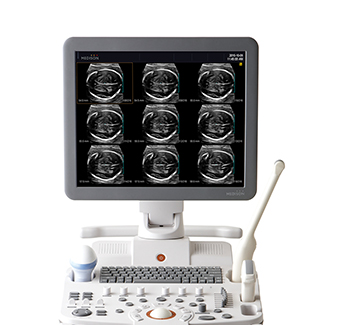
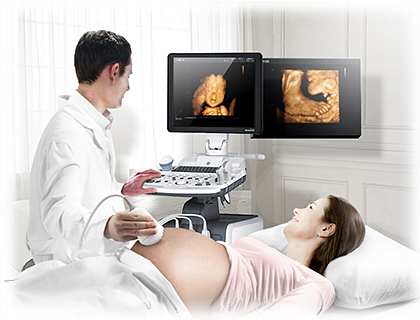
SonoAce R7 enables an increased diagnostic accuracy. By incorporating realistic 4D imaging, this technology delivers optimised results in a variety of applications. This increases user confidence and leads to a more informed decision-making process for medical professionals. The enhanced image quality comes at no expense to the unit’s overall mobility.
Dynamic MR™ / Dynamic MR Plus™
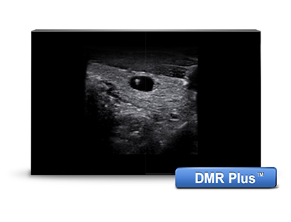
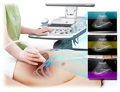
Combining both Object and Pixel Filtering, Dynamic MR™ technology significantly reduces extraneous noise and speckle echoes to create a clearer image of the area in question. This second-stage filtering technique enriches both grey scale and contrast resolution, while also enhancing border detection. Reverberation artifact is thereby reduced, resulting in sharper images for more accurate diagnoses. It assists earlier detection of masses, and is therefore particularly useful when evaluating obstetrical, pelvic and abdominal areas.
Full Spectrum Imaging™ (FSI™)
Regardless of the TX setting, the FSI™ uses the entire radio frequency range and a customised probe to produce more comprehensive and reliable images. Broadening the range enables increased penetration and contrast resolution, ensuring better results for operators by reducing speckle and improving signal-to-noise ratio (SNR). This technology can also be applied to harmonic imaging to provide clinics with a more flexible range of diagnostic tools.
3D eXtended Imaging™ (3D XI™)
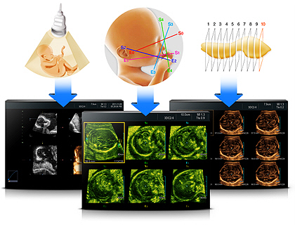
3D eXtended Imaging™ (3D XI™) comprises a suite of three imaging applications to bring cutting edge technology to your organisation. Multi-Slice View™ transforms ultrasound data into a series of sequential images whilst Oblique View™ enables examination of 3D data in a wide range of planes. Finally, the Volume CT™ function allows coronal, sagittal and axial imaging, allowing practitioners to better understand cross-sectional scan information. The combined result is complete control of 3D and 4D volume data manipulation from various perspectives to ensure diagnostic accuracy and confidence.
QuickScan™
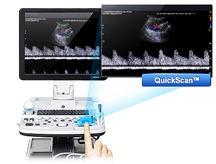
QuickScan™ has been designed to increase workflow efficiency by automatically optimising key imaging parameters at the touch of a button. The resulting increase in efficiency allows your organisation to offer patients an improved service with faster and more accurate diagnoses. This reliable and safe system helps your clinic or hospital to deliver a superior patient experience.
Spatial Compounding Image™ (SCI™)
Spatial Compounding Image™ (SCI™) technology improves signal-to-noise ratio (SNR) and enhances contrast resolution via electronic ultrasound beam steering. This gives operators greater control over their imaging capabilities. SCI™ compounds numerous scan lines to deliver significantly clearer definition in soft tissue planes, with reduced speckle and other interference. The better quality of the detail, compared with Samsung conventional equipment, enables more rapid diagnosis with increased levels of accuracy which leads to increased confidence and a more refined service for your patients.
Enhanced Post-analysis Tool
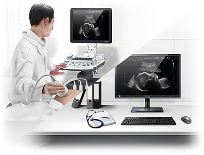
SonoView Pro™ software is optimised to read a variety of media types and formats, including 2D static images, dynamic cine clips and 3D volume data. This post-analysis tool supports medical professionals by allowing faster and more convenient reviewing of patient studies. It enhances productivity by letting the user view exams while away from the ultrasound system, freeing up the unit so that its throughput is increased.
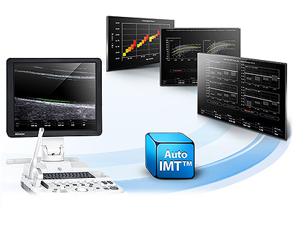
Automated Intima-Media Thickness Measurement™ (Auto IMT™)/strong>
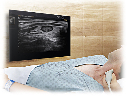
Measurement of the intima-media thickness of the carotid artery wall has become a widely-used technique as it represents a non-invasive, affordable method of diagnosis. Samsung’s Auto IMT™ provides a user-friendly, solution which offers output such as the Framingham Score, risk factors and a user graph. In addition, a comprehensive set of settings including Mean, Max, Standard Deviation and Quality Index measurements are instantly available at the touch of a single button.
User-Customisation
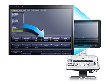
The SonoAce R7 is designed to deliver flexibility and usability for a comprehensive range of clinical situations. The Custom Menu allows the practitioner to preset the items that appear for specific applications according to preference. In this way the options contain only relevant information which enables faster, more efficient operation. In addition, the ability to save edited settings means secure storage and easy retrieval of files, while the Body Marker Edit function allows for clear and simple referencing.
