|
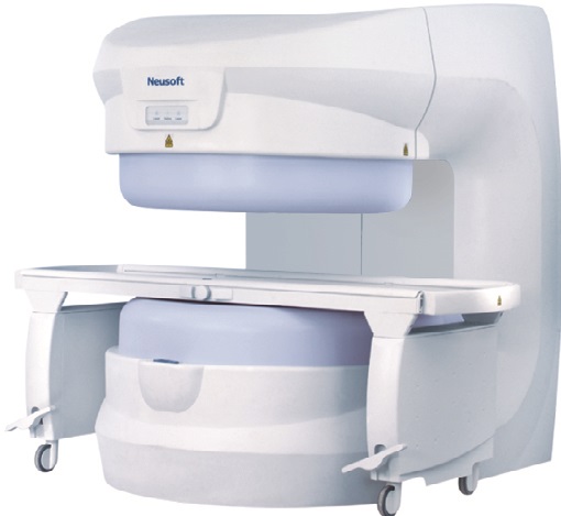
Superstar 0.35T
Image is Everything
Superstar 0.35T MRI system is designed and developed to provide the optimal openness and patient comfort for a wide range of procedures, while providing excellent diagnostic image quality. |
Features
Phased Array Multi-Channel Receiving Coils
|
Clinical Images
| Brain
|
|
|||
 |
 |
 |
||
| Part: brain Plane: axial Weighted and sequence name: T1WI:FFE3D Feature: High SNR and excellent spatial resolution |
Part : brain Plane: axial Weighted and sequence name: T2WI:TSE Feature: High contrast between white matter and grey matter is good |
Part : brain Plane: axial Weighted and sequence name: FLAIR:IR-TSE Feature: High contrast between white matter;and grey matter, the effect of water suppression, a technology artifact from respiration, nearly disappear High SNR and excellent spatial resolution |
||
|
Abdomen and Pelvis
|
||||
 |
 |
 |
||
| Part: upper abdomen Plane: axial Weighted and sequence name: T1WI:TSE Feature: With respiratory gating technology artifact from respiration nearly disappear High SNR and excellent spatial resolution |
Part :upper abdomen Plane: axial Weighted and sequence name: T2WI:TSE Feature: With respiratory gating artifact from respiration nearly disappear High SNR and excellent spatial resolution. Inner structure inside liver revealed clearly |
Part :upper abdomen Plane: axial Weighted and sequence name: T1WI FFE Feature: With breath-hold technology artifact from respiration nearly disappear |
||
|
Spine
|
||||
 |
 |
 |
||
| Part: cervical spine Plane: sagittal Weighted and sequence name: T1WI:TSE Feature: Homogeneity of signal intensity is excellent in a large FOV |
Part: cervical spine Plane: sagittal Weighted and sequence name: T2WI:TSE Feature: Homogeneity of signal intensity is excellent in a large FOV. Motion artifacts are eliminated and there is high contrast between spinal cord and CSF. |
Part : cervical spine Plane: axial Weighted and sequence name: BFFE3D steady state Feature: High contrast between spinal cord and CSF. Clear nerve root display illustrates excellent imaging resolution. |
||
|
Extremity and Join
|
||||
 |
 |
 |
||
| Part: knee joint Plane: sagittal Weighted and sequence name: T1WI:TSE Feature: High SNR and excellent spatial resolution |
Part: knee joint Plane: coronal Weighted and sequence name: T2 STAR:FFE Feature: The sequence is especially useful to observe the cartilage. Bone, cartilage and meniscus are clearly differentiated. |
Part: hip joint Plane: coronal Weighted and sequence name: T1WI:TSE Feature: High SNR and excellent spatial resolution |
||
 |
||||
| Part: hip joint Plane: coronal Weighted and sequence name: FS: IR-TSE Feature: Scan time is short for this proton density fat suppression sequence. |
||||
|
Clinical Technology
|
||||
 |
 |
 |
||
| Part: willis circle Plane: A_P view Weighted and sequence name: FFE3D Feature: MRA technology includes: Slinky acquisition, Partial Echo Acquisition, TOF, and Flow Compensation.The artery is displayed continuously without shutter artifacts |
Part: carotid vertebral artery Plane: A-P view Weighted and sequence name: FFE2D Feature: MRA technology includes:Slinky acquisition, Partial Echo Acquisition, TOF, and Flow Compensation. |
Part : abdomen MRCP Plane: coronal A-P view Weighted and sequence name: Heavy T2WI TSE-3D Feature: The small bile duct inside and outside liver is revealed clearly. Motion artifact nearly disappear with the respiratory gating technology |
||
 |
||||
| Part: inner ear Plane: A_P view Weighted and sequence name: B-FFE3D Feature: This MIP image of inner ear clearly displays three semicircular ducts. |


